Allergic Rhinitis Presentation Template

Allergic Rhinitis: Medical PowerPoint Template
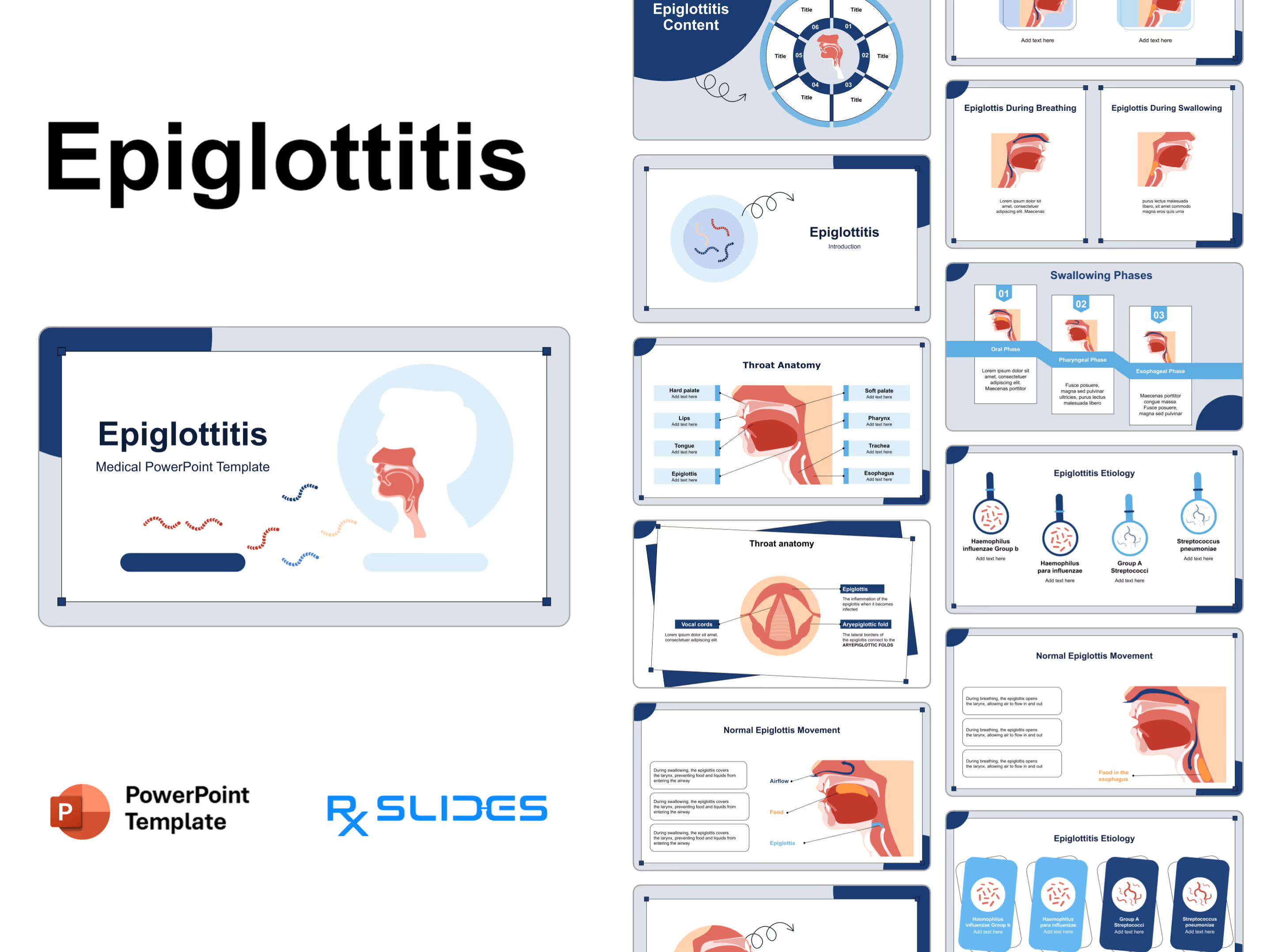
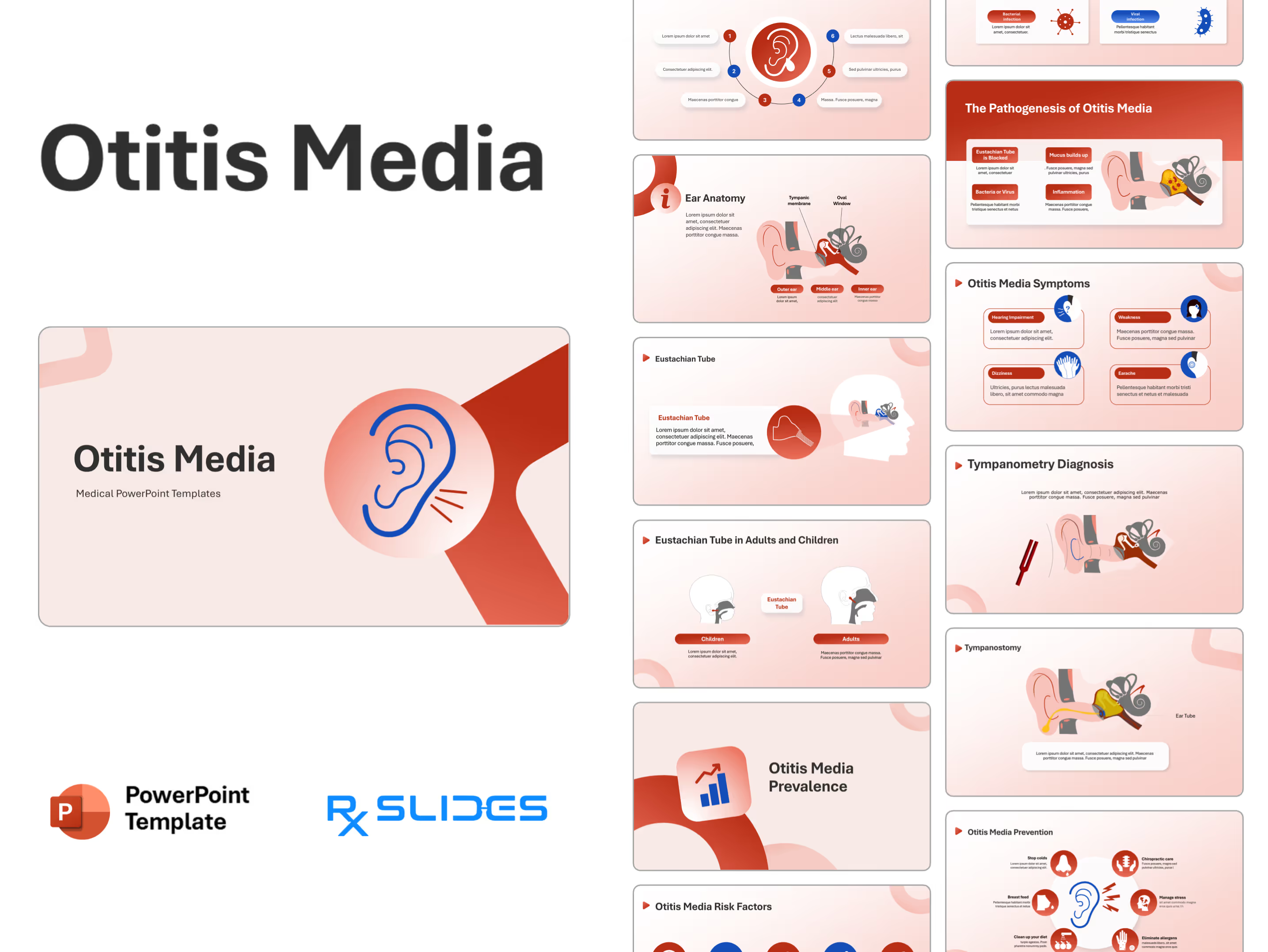
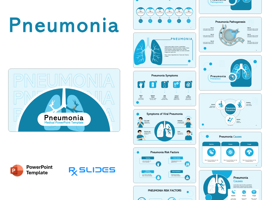
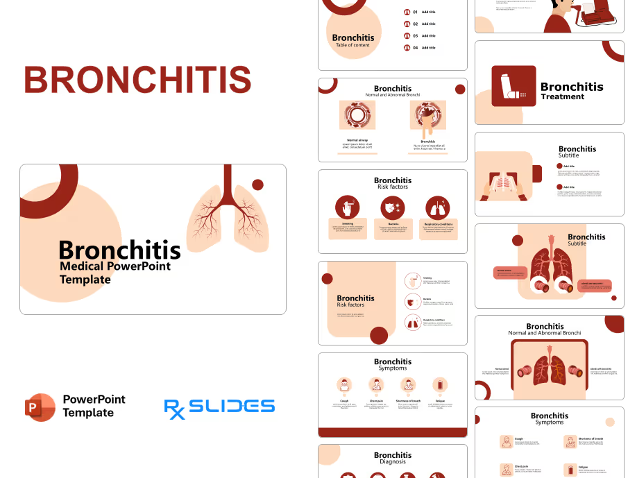
- The Allergic Rhinitis PPT template is an animated medical PowerPoint template that will help you realize the full potential of your presentation.
- The RxSlides template includes medical animations and infographics, which will attract your audience.
- You can rely on our demonstrated infographics to give your audience a dynamic and appealing allergic rhinitis presentation.
- You can explore more related presentations such as the fexofenadine PowerPoint template.
- Suppose you need more templates to explain lung anatomy, gas exchange, and respiratory diseases. In that case, our Respiratory System PowerPoint Templates offer a solution with pre-designed slides featuring high-quality diagrams and animations.
Allergic Rhinitis PowerPoint Template Content
- Creatively designed cover slides to start your Allergic Rhinitis PPT presentation with.

- Editable, customized table of contents that will help you explain the contents of your Allergic Rhinitis PPT presentation.

Allergic rhinitis definition
- Allergic Rhinitis (AR) is an allergic disease, an atopic disease characterized by symptoms of nasal congestion, clear rhinorrhea, sneezing, postnasal drip and nasal pruritis.

Allergic rhinitis prevalence
- The prevalence of Allergic Rhinitis on global and local levels is visualized on an infographic; you can change the percentage and country of the prevalence according to your data, which makes the prevalence data easily described.

Allergic rhinitis risk factors
- Allergic rhinitis risk factors are explained with related medical icons, including smoking, asthma, early fed infants, genetic traits, positive family history, allergen-specific IgE >100 IU/ml serum IgE and male gender.

Allergic rhinitis causes
- Causes are explained with medical icons.
- Outdoor allergens (such as weed blossoms and pollen, which worsen in spring, summer and early autumn).
- Indoor allergens (such as pet skin crumbs and dust mites that aggravate in winter).
- Mold spores, pet dander, pollen, dust mites and cockroaches.

Allergic rhinitis pathogenesis Slides with animation
- Allergic Rhinitis pathogenesis animated illustrations show how pollen enters the nose. Pollen is taken up by dendritic cells, a type of immune cell that phagocytizes.
- Foreign matter: dendritic cells engulf the pollen granules and present them to lymphocytes called T cells, which are then activated and produce cytokines.
- After the cytokines stimulate B cells, another group of lymphocytes produces IgE antibodies.
- IgE antibodies are released into the blood and bind once IgE binds the mast cells; the mast cells are "prepared" for future re-entry of pollen into the body.
- The mast cells degranulate and release their histamine into the local tissue.
- Histamine causes the capillaries to dilate and become leaky, bringing more fluid and immune cells to the area.
- The pathogenesis manifests itself physically, as histamine causes inflammation and swelling of the nasal mucosa, which leads to excessive mucus production and nasal drip. Excessive mucus can block two fundamental opening structures in the nose, such as the nasolacrimal duct, causing tearing of the eyes and the eustachian tubes, causing a feeling of plugged ears.
- The nerves in the nasal passages can also become irritated and cause sneezing.

Allergic rhinitis classification
- Allergic Rhinitis is classified into:
- Seasonal Allergic Rhinitis (occurs in spring, summer and early autumn when trees and weeds are blooming and pollen counts are higher)
- Perennial allergic Rhinitis (can happen year-round).
- Both are provided with fully customizable medical illustrations.

Allergic rhinitis symptoms
- Allergic Rhinitis signs and symptoms are explained with editable visuals of sneezing, runny nose, nasal congestion, sore throat, headache, sinus pain, earplugs, eyes dark circles and trouble breathing.

Allergic rhinitis diagnosis
- Diagnostic methods are carefully designed using animated editable icons and illustrations, including physical examination, general evaluation, medical history, blood test, skin prick test and nasal smear.
- The skin prick test is well explained using animation showing samples of different allergens placed on the skin; the skin is scratched or pricked with a needle, allowing allergens to penetrate the surface; in case of allergy, the area will show redness, itching and irritation between 15 and 30. After a few minutes, raised, hive-shaped welts called wheals are developed.

Treatment options for allergic rhinitis
- Treatment options are visualized and explained using editable illustrations and icons in the Allergic Rhinitis PPT, making it presented more efficiently, including:
- Antihistamines (such as loratadine, cetirizine, fexofenadine and levocetirizine), the mechanism of action is explained using animated illustrations that show how Antihistamines bind to H1 receptors, preventing histamine from binding and shutting it down the in a signal that in causes allergic in symptoms cascade.
- Decongestants (pseudoephedrine, oxymetazoline and phenylephrine): animated illustrations explain that decongestants activate alpha-adrenergic receptors in blood vessels and cause blood vessels to constrict.
- Corticosteroids (beclomethasone dipropionate, budesonide, flunisolide, mometasone propionate furoate and triamcinolone acetonide).
- The animated slides explain how leukotriene inhibitors or antagonists (such as montelukast) bind and block leukotriene receptors, inhibiting leukotrienes from binding to their receptors.
- Immunotherapy and nasal irrigation.

Prevention of allergic rhinitis
- Prevention includes healthy lifestyle choices such as wearing sunglasses, using hypoallergenic bedding and covers, washing bedding regularly, using a vacuum with a HEPA filter, washing pets at least once every two weeks and not allowing pets in bedrooms.

RxSlides visuals for allergic Rhinitis PowerPoint presentation
- A Set of PowerPoint icons and illustrations related to Allergic Rhinitis disease will help you customize the content of this 100% editable presentation according to your content and audience interest.

Features of the Template
- 100% editable PowerPoint template.
- Editable colors, you can change according to your presentation style and company branding guidelines.


