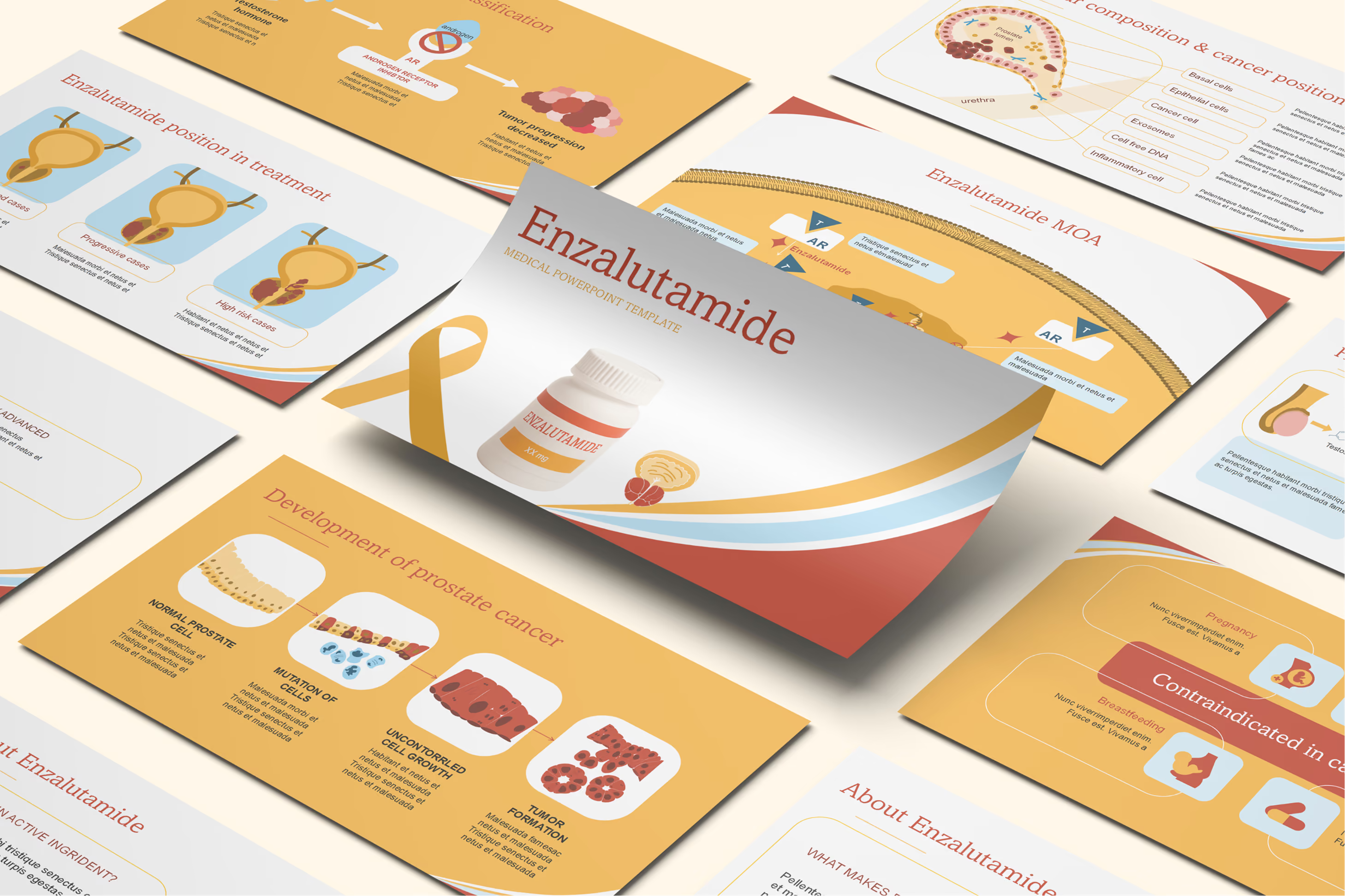
Retinopathy PowerPoint Template

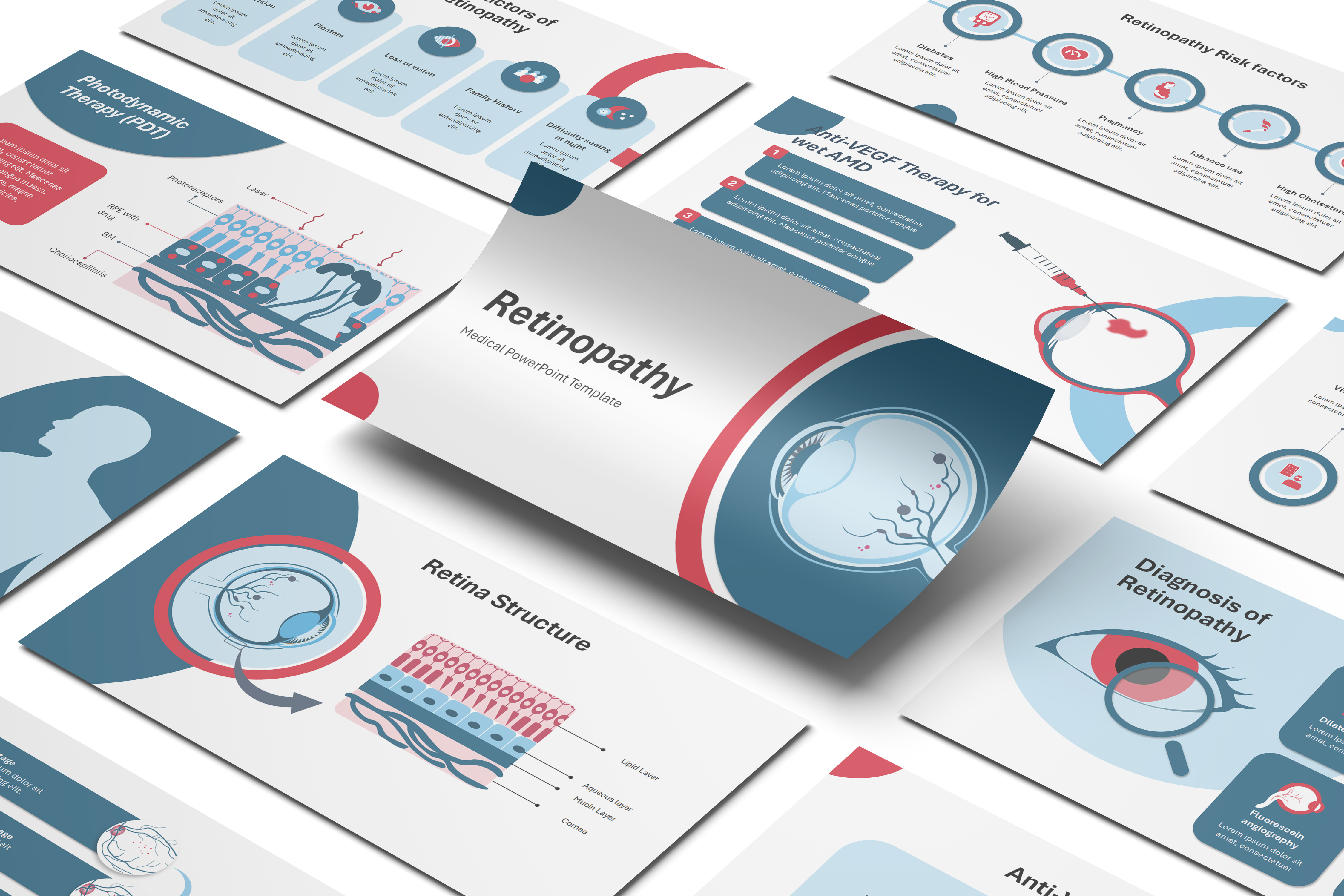




Template Preview
Designed for healthcare professionals creating clear and structured medical presentations.
Ideal for doctors, medical educators, and pharmaceutical teams preparing lectures, clinical education, product presentations, and training.
Fully editable in PowerPoint, allowing you to customize diagrams, charts, and slide layouts with your own medical content
Ready-to-use presentation structure that saves time and helps you build professional healthcare presentations quickly.
Fully Animated
Built-in animations help you present medical information clearly and make complex concepts easier to understand.
Editable Colors and Shapes
Editable colors and shapes help you easily customize the slides to match your content and presentation style, just drag and drop your content.
Professional medical layouts
Professional medical layouts help you organize clinical information, medical data, and treatment insights in a clear and structured way.

Created by Medical Professionals
This template prioritizes medical accuracy, clarity, and real-world usability. Each slide is designed with a medical audience in mind, helping you present clinical information, data, and treatment insights in a way that is structured, credible, and easy for healthcare professionals to understand.
Medical Training for Pharmaceutical & Life Sciences Companies
Medical Affairs and Training teams building ophthalmology onboarding content can use this Retinopathy template to cover the full range of retinal disease beyond diabetes alone, including hypertensive retinopathy, retinal vein occlusion, and medication-related causes, alongside the more familiar NPDR-to-PDR progression. New ophthalmology sales trainees and Medical Science Liaisons get a visual foundation for fundoscopy findings, OCT and fluorescein angiography interpretation, and treatment options spanning laser photocoagulation, anti-VEGF therapy, and vitrectomy. Because the template's cause section extends past diabetes to macular degeneration and glaucoma-related changes, trainers supporting a broader retinal-disease portfolio can use one resource instead of stitching together disease-specific materials. New hires finish training able to place a given retinal finding within its full range of possible causes.

Medical Congress & Scientific Conference Presentations
Ophthalmologists, retina specialists, and endocrinologists preparing a conference talk on retinal disease can adapt this template's illustrated eye anatomy and pathogenesis slides rather than assembling separate diagrams for each retinopathy subtype. The side-by-side comparison of proliferative and non-proliferative mechanisms gives a speaker a ready teaching sequence, while the multi-cause content, spanning diabetes, hypertension, medications, and vascular occlusion, supports a broader differential-diagnosis discussion than a diabetes-only presentation would allow. A Key Opinion Leader speaking at an ophthalmology or endocrinology meeting can reuse the animated illustrations across sessions while swapping in institution-specific prevalence data. The template's diagnostic and treatment sections keep the technical discussion current for a specialist audience.

Pharmaceutical Product & Medical Affairs Presentations
Medical Affairs teams supporting an anti-VEGF or ophthalmology product can use this Retinopathy presentation as broader disease-state groundwork for scientific exchange, covering retinal pathophysiology across multiple causes before narrowing to a specific etiology relevant to the product in question. Medical Science Liaisons can deliver the customized material at advisory boards or KOL discussions to establish shared clinical context, referencing fundoscopy and OCT findings before any product-specific efficacy data enters the conversation. Because the presentation stays disease-focused rather than promotional, it functions as a starting point still subject to the customer's own medical and compliance review before any field use. Ophthalmology and retina teams gain a scientific reference broad enough to cover diabetic, hypertensive, and other retinopathy etiologies.

eLearning & SCORM Training Modules
Learning & Development teams training new ophthalmology technicians or optometry students can customize this Retinopathy template with institution-specific protocols, then convert the finished presentation into a SCORM-compliant module for their learning management system when self-paced study fits the program better. The animated pathogenesis and diagnostic-imaging slides translate naturally into interactive checkpoints within an e-learning course covering multiple retinopathy causes. Where a program needs a narrower focus, such as diabetic retinopathy alone or a version tailored for optometry versus ophthalmology learners, the RxSlides team can adjust the template's scope and branding. A fully custom presentation can also be designed from scratch when no existing template matches the specific curriculum.
Medical & Healthcare Education
Medical and optometry faculty teaching retinal disease can use this template to walk students through eye anatomy and the range of conditions that cause retinopathy, from diabetes and hypertension to retinal vein occlusion and macular degeneration, without assembling separate lecture materials for each cause. The proliferative-versus-non-proliferative comparison gives students a clear framework distinct from a diabetes-only presentation, useful for teaching differential diagnosis in a retinal disease unit. Diagnostic content covering fundoscopy, OCT, and fluorescein angiography connects the underlying pathology to procedures students will observe in ophthalmology or optometry rotations. Students leave with a broader view of retinopathy that extends beyond the diabetic-specific cases they may already be familiar with.
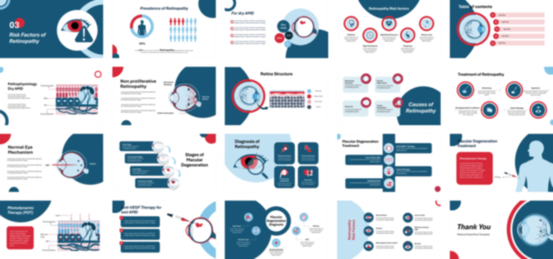
The Retinopathy PowerPoint template opens with cover slides and an editable table of contents before dynamic illustrations detail eye anatomy, including the lens, vitreous body, optic nerve, cornea, pupil, and ciliary body in a set of editable medical diagrams. An infographic with changeable percentages then visualizes the prevalence of retinopathy on global and local levels.
Animated medical icons illustrate risk factors including diabetes, high blood pressure, high cholesterol, pregnancy, and tobacco, noting that diabetic retinopathy specifically stems from high blood sugar. Medical visuals then explain further causes such as certain medications, retinal vein occlusion, macular degeneration, and glaucoma.
Two animated illustrations explain the distinct mechanisms behind proliferative and non-proliferative retinopathy. Establishing this mechanism first gives educators a shared reference point before medical icons depict symptoms including blurred vision, loss of vision, floaters, difficulty seeing at night, dark spots, and eye pain.
The presentation covers diagnostic methods including visual acuity tests, dilated eye exams, fluorescein angiography, and tonometry before animated illustrations detail treatment options such as vitrectomy, injections, photocoagulation, and laser treatment. It closes with preventive methods including regular eye exams and a healthy diet, alongside complications such as vision loss, retinal detachment, glaucoma, and neovascularization, illustrated in a set of editable medical icons and illustrations.
Ready To Transform Your Presentation ?
Pay once, use forever