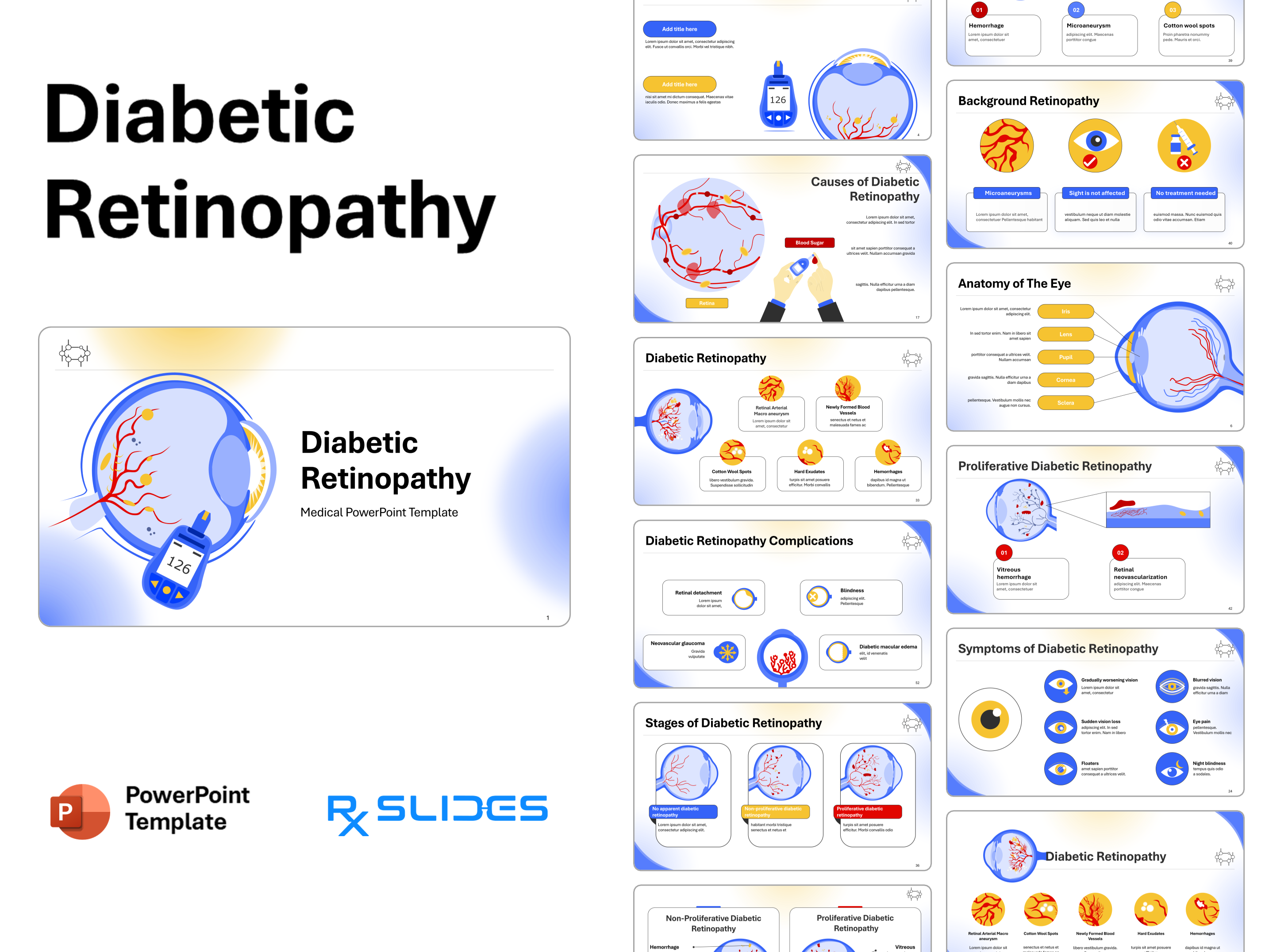
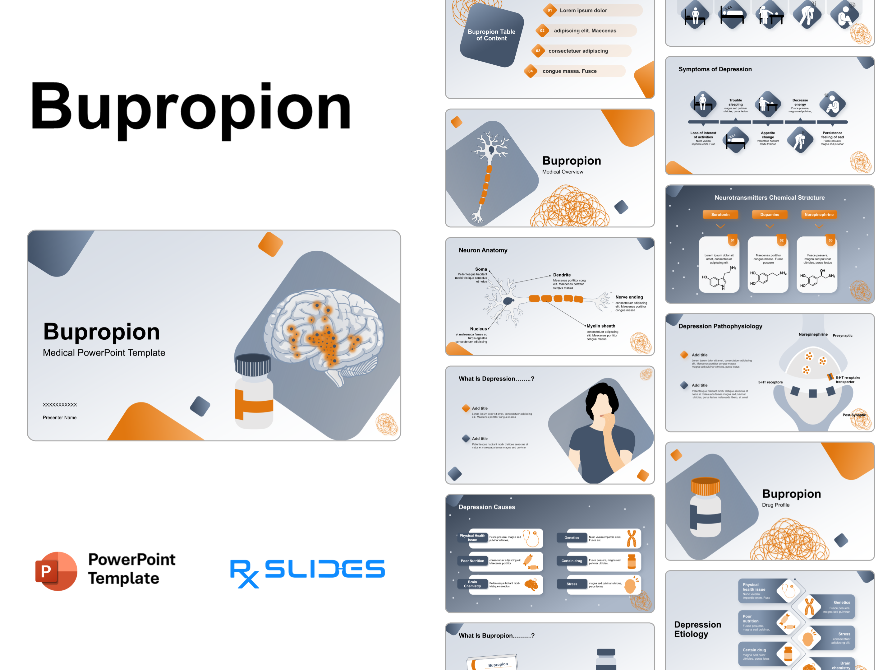
Age-related Macular Degeneration PPT Template


No items found.
Age-Related Macular Degeneration Presentation: Medical PowerPoint Template
- Age-related macular Degeneration (ARMD)PPT template is a dynamic medical PowerPoint template that will help you realize the full potential of your presentation. The RxSlides animated template includes medical animations and infographics, which will attract your audience.
- You can rely on our demonstrated infographics to provide your audience with a dynamic and appealing macular degeneration presentation.
- These animated slides are valuable for ophthalmologists, medical students and healthcare workers and can be used for patient education purposes.
- Our Eye Disease PowerPoint Templates provide a framework for impactful presentations with dynamic animations and clear illustrations that convey eye disease pathogenesis and treatment procedures.
Age-Related Macular Degeneration PowerPoint Template Content
- Nice-looking cover slides to start your age-related macular degeneration PPT presentation with.

- An editable table of contents that will help you explain the contents of your age-related macular degeneration PPT presentation.
Anatomy of the eye
- Illustrated anatomy explaining the eye including cornea, lens, ciliary body, vitreous body and macula optical nerve.
Structure of the retina
- The structure of the retina is illustrated, showing that the cells subdivide into three basic cell types: photoreceptor cells, neuronal cells and glial cells.
- The retinal structure is illustrated as layers containing photoreceptors, RPE, BM and choriocapillaris.

Age-related macular degeneration definition
- Age-related macular degeneration (ARMD)is an eye disease that can blur central vision. It happens when aging causes damage to the macula.
Epidemiology with charts and infographics for macular degeneration
- The epidemiology of macular degeneration on global and local levels is visualized in an infographic; you can change the percentage of the prevalence according to your data.

Risk factors with medical icons macular degeneration
- Macular degeneration's most common risk factors include smoking, age, genetics, family history and female gender.

Comparative illustrations between types of macular degeneration
- Animated illustrations demonstrate macular degeneration types as follows:
1. Dry (atrophic) macular degeneration
- Animated illustrations explain that it develops when tiny yellow protein deposits called drusen form under your macula. The built-up deposits dry and thin your macula.
2. Wet (exudative) macular degeneration
- Animated illustrations demonstrate that wet (exudative) macular degeneration occurs when abnormal blood vessels develop under your retina and macula. The blood vessels leak blood and fluids.

Macular degeneration symptoms explained by medical visuals
- Signs and symptoms are explained with visuals of distorted lines, blurred central vision, a need for brighter light, dark patches in central vision and hard-to-distinguish faces.

Animated illustrations show the progression of macular degeneration disease
- Macular degeneration stages are illustrated with animated illustrations demonstrating early, intermediate, late and neovascular.

Diagnostic methods for macular degeneration
- Diagnosis of macular degeneration: the carefully designed PowerPoint slides showing insights into the diagnosis of macular degeneration are eye exam, oct, medical history, Amsler grid test and genetic testing.

Illustrated treatment options for macular degeneration
- Treatment options for macular degeneration are visualized and explained with animated illustrations, including anti-VEGF therapy, low vision aids, laser therapy and supplements. You can add or remove the treatment techniques in this section according to the latest updates.

Preventive methods of macular degeneration
- Macular degeneration prevention includes a healthy diet, exercising regularly, managing health conditions and supplements, quitting smoking and wearing sunglasses.

Complications of macular degeneration
- Complications include blindness, anxiety, social isolation and an increased risk of falls.

RxSlides visuals of the ARMD PowerPoint presentation
- End your set of PowerPoint icons and illustrations related to macular degeneration, which will help you customize the content of this 100% editable presentation according to your content and audience interest.

Features of the Template
- 100% editable PowerPoint template.
- Editable colors, you can change according to your presentation style and company branding guidelines.